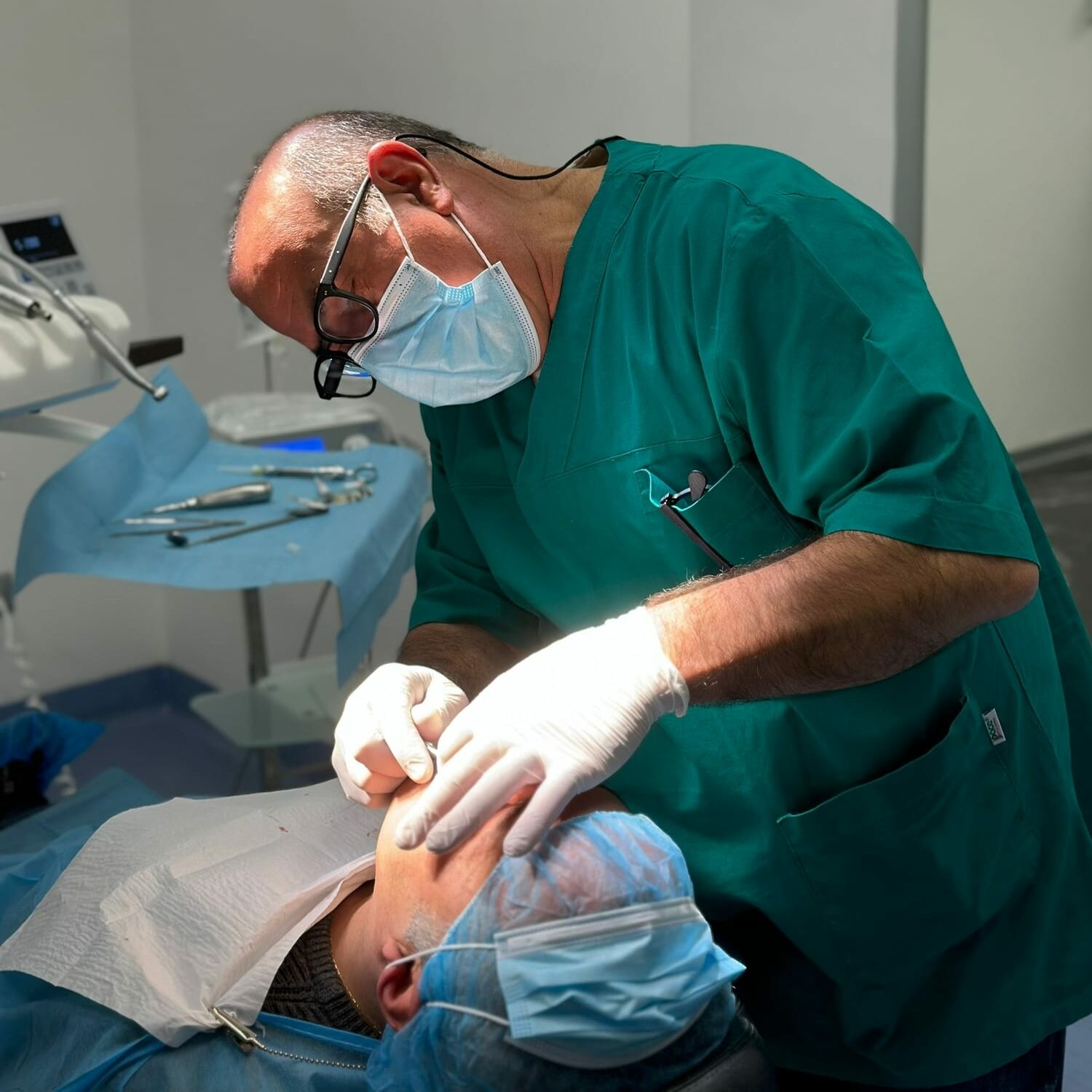
The suitability of a patient to face an implantology intervention is assessed by the dentist on the basis of many factors but two are the main ones: the quantity and quality of the bone.
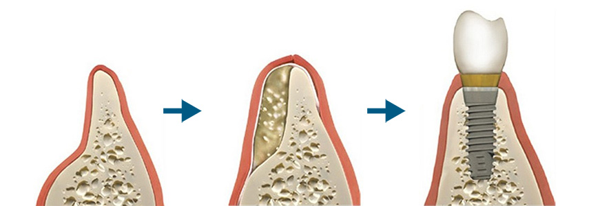
Cases of patients with alveolar bone so small (in height and width) that it is difficult to insert a dental implant are not uncommon. How to solve this problem? There is bone reconstructive surgery, let’s see better what it is.
Bone atrophies and solutions
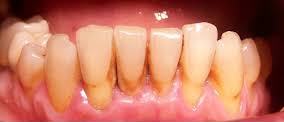
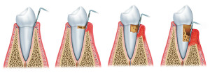
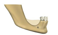 “Bone atrophy” which can be traced back to different causes: when a patient is affected by partial or total edentulism, chewing or eating can be activities that, in the long run, affect the bone reducing its thickness; gingival inflammation can result in periodontitis which, in the more advanced stages, turns into pyorrhea with loss of teeth.
At that point, the implantologist will have to work on the bone by increasing the volume. The reconstructive bone surgery can take place: simultaneously with the insertion of the implant (called peri-implant bone regeneration) or before carrying out implant surgery (i.e. pre-implant bone regeneration).
There are four methods of treatment to correct reduced bone thickness:
- guided bone regeneration (GBR)
- block graft (intra or extraoral bone)
- crest expansion (Split Crest)
- maxillary sinus lift
Guided bone regeneration
The guided bone regeneration treatment (GBR) takes place simultaneously with the implant insertion and consists in the use of resorbable membranes and filling materials. The best material to rely on for the volumetric thickness increase is obviously the patient’s bone, also called autologous bone, there are also synthetic materials that are usually used for smaller fillings.
Block coupling
It involves the removal of bone from intraoral sites, chin or jaw, or from extraoral sites, hip or skull, in the case of very large grafts. But why block graft? The bone taken in shaped blocks is fixed in the receiving site through small titanium screws (trans-cortical screws).
Split Crest
For the horizontal increase of the edentulous ridges that present bone atrophy there is the “Split technique

Crest”. In fact, horizontal bone resorption is one of the most common constraints on implant positioning.
The treatment, which can take place simultaneously with the insertion of the implants, consists in the expansion of the edentulous bone crest with preventive cutting of the bone parallel to the progress of the teeth (sagittal osteotomy).
It is normally adopted for the upper arch (here the bone thickness tends to be greater), when two or more dental elements are missing and the thickness of the residual ridge has a minimum thickness of 3 mm. In the presence of anatomical hourglass or narrow base forms with thinner thicknesses, the Split Crest is contraindicated: autologous bone grafting and the insertion of dental implants in a phase following healing of the grafted site will be preferred.
Sinus lift
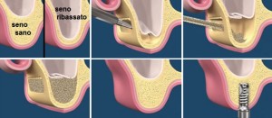
The air cavity located in the posterior area of the upper jaw is called the maxillary sinus. Precisely this anatomical area of the jaw, after the loss of the teeth, undergoes a reduction in height with reabsorption of the bone.
To raise the bone in this precise location, two techniques will be used:
- mini maxillary sinus lift (internal elevation) for an acquisition of bone height of 2-3 mm, when the bone thickness is 6-8 mm;
- large elevation of the maxillary sinus (external elevation) when the height of residual bone in the posterior areas of the jaw is very small and makes positioning of the implants impossible. Filling with autologous particulate bone and / or artificial bone is used.


